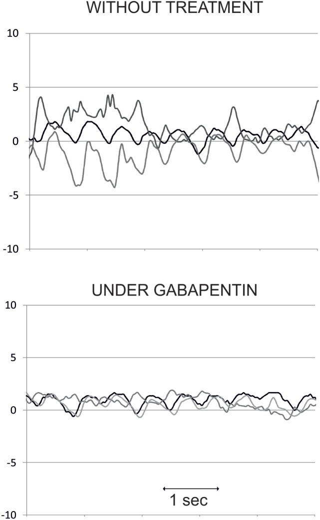
Figure 7.

Eye position (in degrees) traces over time (in seconds) in one oculopalatal tremor patient, without treatment (upper panel) and under gabapentin (lower panel). Dark line: horizontal position, gray line: vertical position, and light gray line: torsional position. Note the decrease in nystagmus amplitude, mainly in the torsional plane, under gabapentin.
