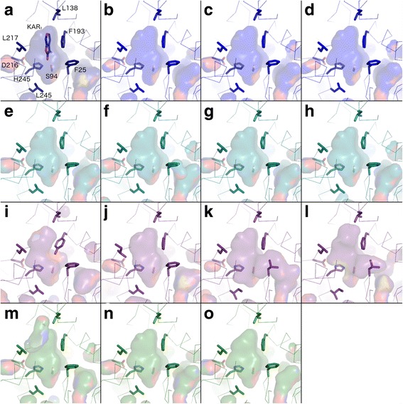
Fig. 6.

Homology models of KAI2 sequences. Models are shown in ribbon representation with the residues that influence the active site cavity shown in stick representation. Cavities are depicted as a transparent surface. Oxygen, nitrogen and sulphur atoms are coloured red, blue and yellow respectively. a The crystal structure of Arabidopsis thaliana KAI2 in complex with karrikin (KAR 1) is shown in navy blue (PDB code 4JYM). Residue numbers correspond to the unified numbering scheme as in Fig. 5; they are –1 relative to those of A. thaliana KAI2. b–d Liverwort KAI2A homology models are shown in royal blue; b Lejeuneaceae sp. c Lunularia cruciata, d Ptilidium pulcherrimum. e–h Liverwort KAI2B models are shown in turquoise; e Riccia berychiana, f Calypogeia fissa, g Lunularia cruciata, h Marchantia polymorpha. i–l Charophyte KAI2 models are shown in purple; i Klebsormidium subtile, j Chara vulgaris, k Coleochaete scutata, l Coleochaete irregularis. m–o Moss KAI2E/F models are shown in green; m Sphagnum recurvatum KAI2E, n Timmia austriaca KAI2F, o Tetraphis pellucida KAI2F
