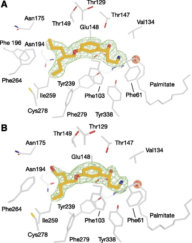
Fig. 3.

Binding of MB-004 and MB-008 to the RPE65 active-site pocket. MB-004 (A) and MB-008 (B) are shown as orange sticks. Residues within 4.5 Å of the MB ligands are shown as gray sticks. The iron cofactor and the iron-bound palmitate ligand are shown as brown spheres and gray sticks, respectively. The green mesh represents a σA-weighted Fo-Fc electron density map calculated after deletion of the ligand and 30 cycles of REFMAC refinement. Residues within 4.5 Å of the ligand were harmonically restrained to their initial positions during refinement. The electron density suggested that the R,S form of MB-008, which was added to the protein as a racemic mixture, was the dominant isomer occupying the binding pocket.
