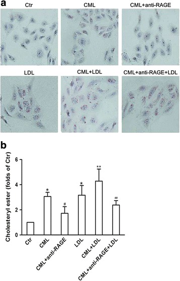
Fig. 3.

Visualization of LDL uptake and lipid droplets in human renal tubular epithelial cell line (HK-2) after Nε -(carboxymethyl) lysine (CML) treatment. HK-2 cells were incubated for 24 h in experimental medium, or medium containing 50 μg/ml CML or 200 μg/ml LDL, or 50 μg/ml CML plus 10 μg/ml anti-RAGE, or 50 μg/ml CML plus 200 μg/ml LDL, or 50 μg/ml CML plus 200 μg/ml LDL and 10 μg/ml anti-RAGE. a Cells were examined for lipid inclusions by Oil Red O staining. The results are typical of those observed in 3 separate experiments (×200). b The concentration of cholesterol ester in HK-2cellswas measured as described in Materials and Methods. Values are mean ± SD of duplicate wells from 3 experiments. *P < 0.05 vs. Ctr; **P < 0.05 vs. LDL group; # P < 0.05 vs. CML group; ## P < 0.05 vs. CML+ LDL group
