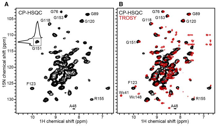
Fig. 6.
2D 1H–15N correlation spectra of Ail in phospholipid bilayers. a Solid-state NMR 1H-detected CP-HSQC spectrum of (u-15N, u-13C, f-2H) Ail in liposomes, recorded at 900 MHz, 30 °C, with 160 scans and a MAS rate of 60 kHz. b Solution NMR 1H-detected TROSY-HSQC spectrum (red) of (u-15N, u-13C, u-2H) Ail in nanodiscs prepared with 2H labeled lipids, recorded at 800 MHz, 45 °C, with 128 scans. The solid-state NMR CP-HSQC spectrum (black) is superimposed

