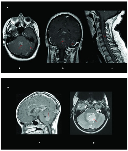
Figure 1. Imaging of pediatric and adult medulloblastomas.
( A) Magnetic resonance imaging of an adult woman who has medulloblastoma in the brain and spine with leptomeningeal spread: ( a) axial T1 of the brain post-gadolinium contrast; ( b) coronal T1 of the brain post-gadolinium contrast; ( c) sagittal T1 of the cervical spine post-gadolinium contrast. ( B) Brain magnetic resonance imaging of pediatric medulloblastomas: ( a) sagittal post-gadolinium WNT tumor; ( b) axial T2 of a SHH tumor. Red arrows delineate the tumor/leptomeningeal disease.

