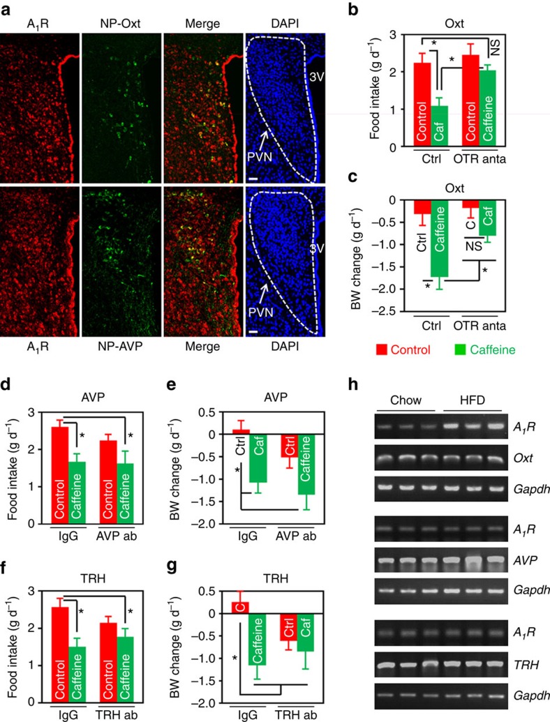
Figure 6. Oxt mediates caffeine's effect on energy balance in the DIO mice.
(a) Double immunofluorescence staining of A1R (red) and Neurophysin I (NP-Oxt, green) or Neurophysin II (NP-AVP, green), which is co-expressed with Oxt or AVP in the PVN, respectively. Cell nuclei were counterstained with DAPI (blue). 3V, third ventricle. Scale bar, 50 μm. (b–g) HFD-fed mice were i.c.v. administered aCSF or IgG as control (Ctrl), and 2 μg of Oxt receptor (OTR) antagonist (anta), L-368,899 (b,c), or 0.5 μg of antibody against AVP (d,e) or TRH (f,g). An hour later, mice were i.c.v. injected control or 10 μg of caffeine (Caf or C). Twenty-four hours food intake (b,d,f) and body weight change (c,e,g) were then measured. ab, antibody. In OTR antagonist experiment, n=10 (Ctrl+Ctrl), 11 (Ctrl+Caf), 9 (OTR anta+Ctrl), 13 (OTR anta+Caf); AVP antibody, n=11 (IgG+Ctrl), 9 (IgG+Caf), 7 (AVP ab+Ctrl), 14 (AVP ab+Caf); TRH antibody, n=7 (IgG+Ctrl), 7 (IgG+Caf), 8 (TRH ab+Ctrl), 10 (TRH ab+Caf). (h) Single-cell RT-PCR analysis of A1R expression in Oxt, AVP or TRH-expressing cells isolated from the PVN of chow or HFD-fed mice. Gapdh was used as an internal control. Data are presented as mean±s.e.m. *P<0.05, one-way analysis of variance (ANOVA) with Bonferroni’s (b,d,e,f) or Newman–Keuls (c,g) post hoc test. NS, not significant.

