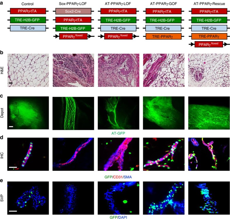
Figure 1. PPARγ regulates APC–blood vessel residency.
(a) Illustration of genetic alleles used to generate: AdipoTrak (AT)-control (PPARγtTA; TRE-H2B-GFP; TRE-Cre); Sox-PPARγ-LOF (Sox2-Cre; PPARγf/tTA; TRE-H2B-GFP); AT-PPARγ-LOF (PPARγf/tTA; TRE-Cre; TRE-H2B-GFP); AT-PPARγ-GOF (PPARγtTA; TRE-H2B-GFP; TRE-PPARγ); and AT-PPARγ-Rescue (PPARγf/tTA; TRE-PPARγ; TRE-Cre; TRE-H2B-GFP). Experiments were performed three times on six mice per group. (b) Representative H&E images from mice described in a. (c) Representative GFP images of freshly isolated subcutaneous IGW depots from mice described in a. Scale bar 10 mm. (d) Representative images of CD31 (endothelial marker) and SMA (mural cell marker) and APC-GFP immunostaining from subcutaneous IGW depots from mice described in a. (e) Representative images of SVPs from subcutaneous adipose depots from mice described in a. Locality of APCs was assessed by GFP fluorescence 12 h after isolation. DAPI was used to visual nuclei and cell number (n=9). Scale bars 100 μm.

