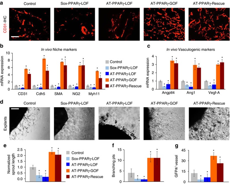
Figure 2. PPARγ is required for WAT niche expansion.
(a) Representative sections from AT-control, Sox-PPARγ-LOF, AT-PPARγ-LOF, AT-PPARγ-GOF and AT-PPARγ-Rescue stained for endothelial marker CD31. (b,c) Quantitative RT–PCR analysis of endothelial and mural cell (nichegenic) markers (b) and vasculogenic genes (c) from total SV cells isolated from the mice described in a. (d–g) Representative images of vascular sprouts of subcutaneous IGW explants from the mice described in a, d. Sprout length (e), branching points (f) and GFP progenitor cell occupancy (g) were quantified. Scale bars 100 μm. Data are means±s.e.m. Experiments were performed three times on eight mice per group. *P<0.05 mutant compared to control levels unpaired t-test, two-tailed.

