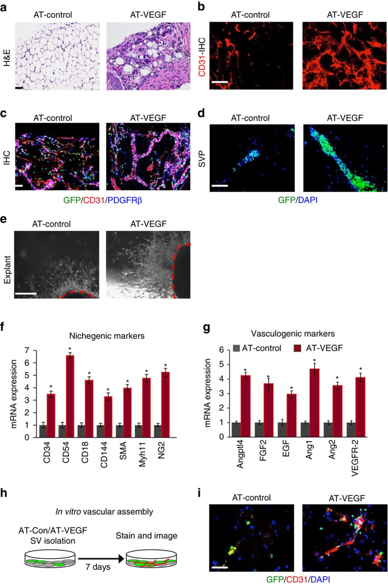
Figure 8. VEGF stimulates niche expansion but blocks fat formation.
(a) Representative images of H&E staining of AT-control (PPARγtTA; TRE-H2B-GFP) and AT-VEGF (PPARγtTA; TRE-VEGF; TRE-H2B-GFP) at 3 months of age. (b) Representative images of CD31 immunostaining of IGW adipose depots from AT-control and AT-VEGF mice. (c) AT-control and AT-VEGF IGW adipose depots were sectioned and stained for GFP, CD31 and SMA. (d) Representative images of SVPs isolated from 3-month old AT-control and AT-VEGF mice and analysed for GFP locality. (e) Representative images of vascular sprouts from subcutaneous IGW adipose explants from 3-month old AT-control and AT-VEGF mice. (f,g) Quantitative RT–PCR analysis of niche and vasculogenic genes from 2-month old AT-control and AT-VEGF mice. (h,i) Total SV cells were isolated from AT-control and AT-VEGF mice (n=6). Cells were cultured for 7 days and then examined for vascular assembly by examining CD31 staining and GFP+ cell co-localization. Scale bars 100 μm. Data are expressed as means±s.e.m. Experiments were performed three times on 6–10 mice per group. Scale bars 100 μm. *P<0.05 unpaired t-test, two tailed: mutant compared to control levels.

