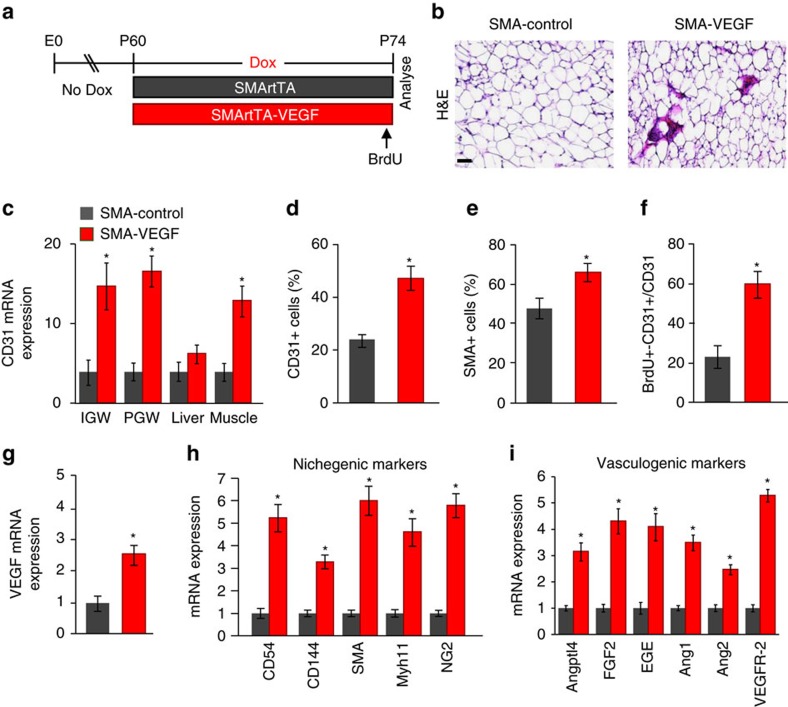
Figure 9. VEGF stimulates niche expansion from adult APCs.
(a) Diagram of experimental paradigm. At P60, SMA-rtTA control or SMA-rtTA; TRE-VEGF (SMArtTA-VEGF) mice were administered Dox for 2 weeks. BrdU was administered 24 h before final analysis. Experiments were performed three times on seven mice per group. (b) Representative images of H&E staining of SMArtTA-control and SMArtTA-VEGF mice described in a. (c) Quantitative RT–PCR analysis of CD31 mRNA expression from denoted tissues. (d–f) Total SV cells were isolated from subcutaneous IGW depots mice described in a. Cells were stained and examined for CD31 (d), SMA (e) or BrdU and CD31-positive cells by flow cytometry. (g–i) Quantitative RT–PCR analysis of VEGF mRNA expression (g), nichegenic (h) and vasculogenic (i) genes from mice described in a. Scale bar 50 μm. Data are expressed as means±s.e.m. *P<0.01 unpaired t-test, two tailed: mutant compared to control levels.

