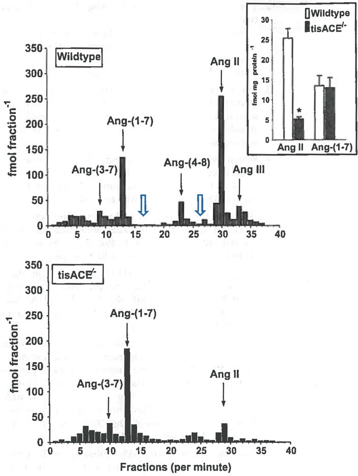
Fig 4.

Combined high-performance liquid chromatography (HPLC) separation and RIA analysis of pooled kidney extracts from wildtype and tissue ACE knockout (tisACE−/−) mice. The collected HPLC fractions were completely evaporated and assayed by the Ang-(1–7) RIA (fractions 1–20) and the Ang Il RIA (fractions 21–40). The HPLC solvent system is 0.1 % HFBA (mobile phase A) and 80% acetonitrile/0.1 % HFBA (mobile phase B). Gradient conditions for Ang-(1–7) and Ang II separation were: 15–40 % B linear over 20 min; 40 % B isocratic for 20 min; 40–15 % B linear for 10 min, 15 % isocratic for 20 min at a flow rate of 0.35 ml/min at 25 °C. The arrows indicate the elution peaks for Ang-(3–7), Ang-(1–7), Ang-(4–8), Ang II, and Ang-(2–8) (Ang III). The open arrows indicate expected elution times for Ang-(2–7) and Ang-(3–8), respectively. Inset intrarenal concentration of Ang II and Ang-(1–7) (fmol/mg protein) in wildtype (n=8) and f/sACE−/−mice (n=8); *P<0.001 versus wildtype. Figure adapted with permission from Modrall et al. [9]
