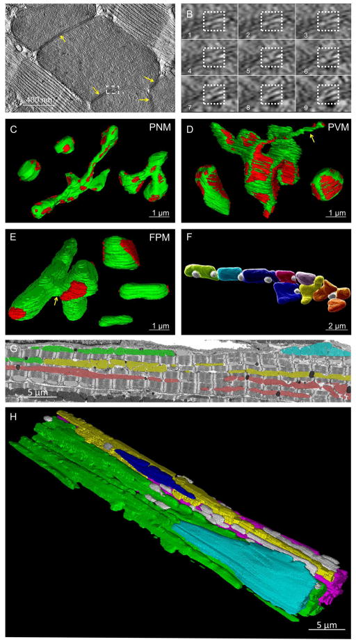
Figure 2. Heart Mitochondrial Morphology and Connectivity.
A) Single IMJ (yellow arrows) tomogram slice. Dashed box shown in B. B) Sequential tomograms of membrane to membrane contact site (dotted box). C–E) Mitochondrial morphologies (green) and locations of IMJ (red). C) Compact (upper left) and Elongated (middle and right) PNM. D) Elongated, Nanotube (yellow arrow), and Compact PVM (left to right). E) Connector (left), Compact (right upper), Elongated (right middle), and Non-connected (right lower) FPM. Yellow arrow – sheet-like connecting structure. F) Adjacent FPM (assorted colors) and lipid droplets (white). G) FIB-SEM frame. Mitochondria in the same color are IMJ-coupled. H) 3D rendering of mitochondrial subnetworks. Each color represents a different IMJ-coupled subnetwork. Grey - not network connected. Represents 5 volumes, 4 animals. See Table S1 and Movie S1.

