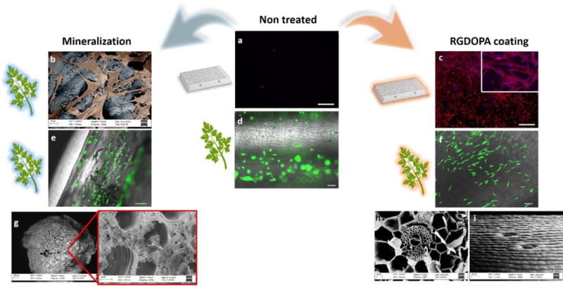
Figure 3. Biofunctionalized plants as scaffolds for human cells.

a, Rhodamine-phalloidine staining of actin filaments (red) and dapi staining of nuclei (blue) of hDFs cultured for 2 days in ultralow attachment polystyrene wells (scalebars 500μm). b, Color-enhanced SEM micrographs displaying hDFs (orange) adhering on a biomineralized parsley stem (blue). c, Rhodamine-phalloidin and DAPI staining of hDFs cultured for 2 days on RGDOPA-coated ultralow attachment polystyrene well (scalebars 500μm). d–f, calcein staining of hDFs seeded on decellularized parsley stems untreated (d), biomineralized (e), and coated with RGDOPA (f) (scalebars 100μm). The images show selective cell adhesion on functionalized surfaces. g, a decellularized parsley stem following 7 days incubation in SBF showing growth of a mineral coating with spheroidal morphology within the pores of the stem. The smallest pores appear to be occluded by the mineral, but larger pores and vascular bundles are open and morphological features are maintained after the mineralization. h–i, SEM micrographs of decellularized stems coated with RGDOPA. A cross-section of a Calathea zebrina stem (h) shows that the RGDOPA coating does not occlude even the smallest (~2 μm) pores; a side-view of a vanilla stem (i) shows that the topographical cues of the stems are still evident after the RGDOPA coating.
