
Figure 1.
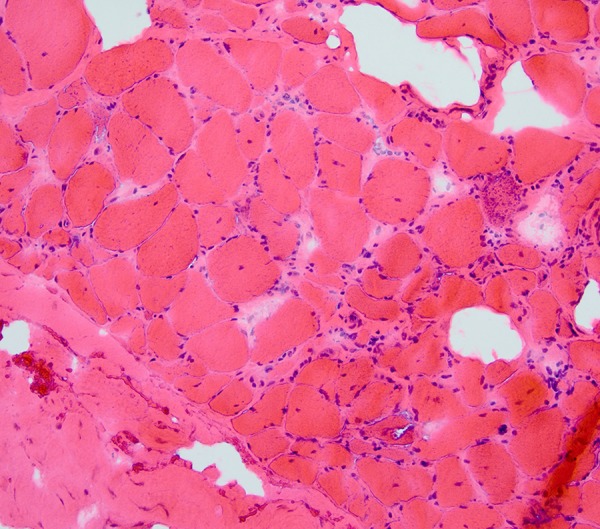
Quadriceps muscle biopsy. Hematoxylin and eosin stain. The muscle fiber size is varied. Many have visible nuclei. Some clusters of atrophied fibers are seen, particularly in the lower right. Endomysial connective tissue is increased in some regions. Inflammation is evident, with lymphocytes. These are most marked in the endomysium. They surround myocytes, which are invaded in some cases (e.g., with splitting of the fiber), as visible in the lower right. No clearly defined inclusion bodies or rimmed vacuoles are noted.