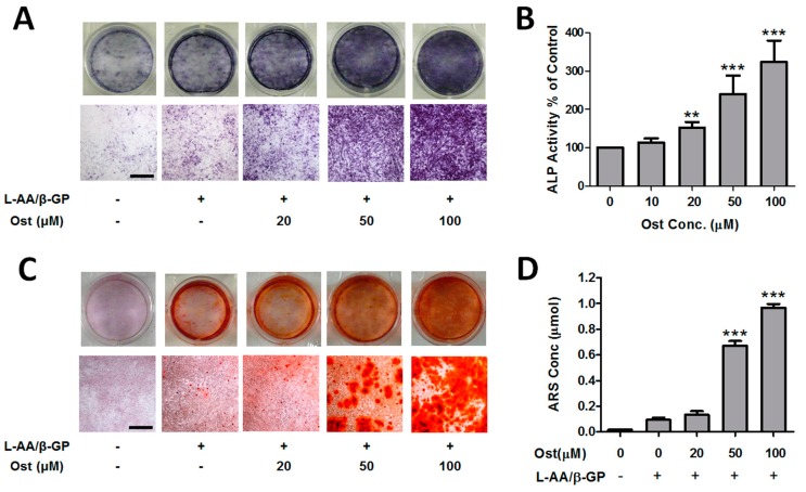
Figure 2.
Osthole promoted osteogenic differentiation in osteoblasts. MC3T3-E1 cells were treated with 0–100 μM osthole in growth or osteogenic medium for 12 (A,B) or 24 days (C,D). (A) Representative macroscopic and microscopic photos of ALP staining cells (n = 4); (B) ALP activities were measured by ALP-AMP kit (n = 6); (C) Representative photos of ARS staining cells (n = 4); (D) cell mineralization was quantified by extraction of ARS dye (n = 4). One-way ANOVA was followed by Tukey’s test and compared to the vehicle control, ** p < 0.01, *** p < 0.001. Bar = 500 μm.

