
Figure 2.
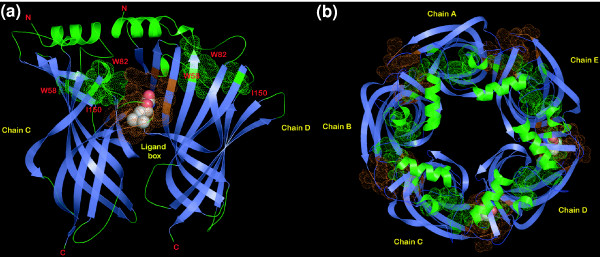
Cartoon representations of ACHB. (a) The ligand-binding dimer of ACHB; (b) the pentamer of ACHB. The representations were derived from the crystal structure of the snail acetylcholine-binding protein (PDB 1UV6). Residues forming the ligand-binding box are shaded orange. The chain of residues that could potentially act as a conduit for transmission of conformational changes is colored green and the prominent conserved ones among them have been labeled.