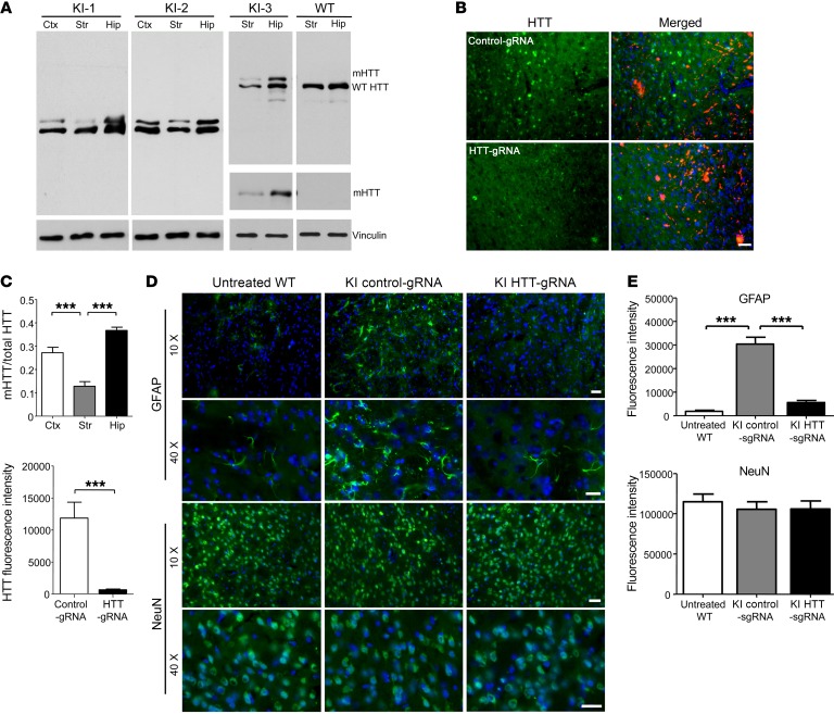
Figure 3. Removal of mHTT in neuronal cells alleviates neuropathology in 13-month-old heterozygous HD140Q-KI mouse striatum.
(A) Western blotting shows the reduction of mHTT in brain tissues from 3 heterozygous HD140Q-KI (KI-1, KI-2, and KI-3) and WT mice. 2166 Antibody was used to show both mHTT and WT HTT. 1C2 Antibody was used to show only mHTT. Replicate samples run on separated blots are presented. (B) Double immunostaining with 1C2 antibody confirmed the depletion of mHTT by AAV-HTT-gRNA. A heterozygous HD140Q-KI mouse injected with AAV-control-gRNA served as a control. Scale bar: 20 μm. (C) Quantitative assessments of the relative ratio of mHTT to total HTT in A (left; n = 8; ***P < 0.001, by 1-way ANOVA with Tukey’s test) and relative levels of mHTT staining in B (right; n = 8; ***P < 0.001, by Student’s t test). (D) Double immunostaining of striatum (from 9-month-old injected mice examined at 13 months of age) shows decreased GFAP levels by HTT-gRNA compared with control-gRNA. There was no difference in NeuN staining. Scale bars: 20 μm. (E) Quantitative assessment of the relative levels of GFAP and NeuN staining (n = 8). The staining intensity for each mouse was the average from three ×10 images. ***P < 0.001, by 1-way ANOVA with Tukey’s test. Data represent the mean ± SEM.

