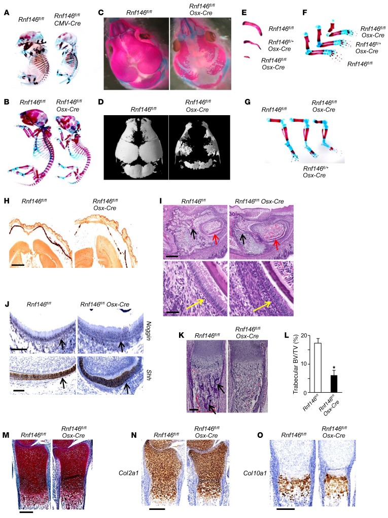
Figure 1. RNF146 deficiency in osteoblasts causes a CCD-like syndrome in mice.
(A) Whole-mount skeletons of Rnf146fl/fl and Rnf146fl/fl CMV-Cre E15.5 embryos stained by alizarin red and Alcian blue. (B and C) Whole-mount skeletons (B) or the calvarium (C) of Rnf146fl/fl and Rnf146fl/fl Osx-Cre newborn pups stained by alizarin red and Alcian blue. (D) μCT reconstruction of the calvarium of Rnf146fl/fl and Rnf146fl/fl Osx-Cre newborn pups. (E–G) Clavicles (E) or limb bones (F and G) of Rnf146fl/fl, Rnf146fl/+ Osx-Cre and Rnf146fl/fl Osx-Cre newborn pups stained by alizarin red and Alcian blue. (H) Alizarin red staining of the calvarium from Rnf146fl/fl and Rnf146fl/fl Osx-Cre newborn pups. Scale bar: 1 mm. The calcified tissue appears red. (I) H&E staining of mandibular incisor from Rnf146fl/fl and Rnf146fl/fl Osx-Cre newborn pups. Scale bars: 300 μm (top panels); 50 μm (bottom panels). Black, red, and yellow arrows indicate periodontal alveolar bone, enamel, and ameloblasts, respectively. (J) ISH of noggin (top panel) and Shh (bottom panel) in mandibular incisor from Rnf146fl/fl and Rnf146fl/fl Osx-Cre newborn pups. Scale bars: 100 μm. Black arrows indicate ameloblasts. (K) H&E staining of tibiae from Rnf146fl/fl and Rnf146fl/fl Osx-Cre newborn pups. Scale bar: 150 μm. Black arrows indicate trabecular bone. (L) Histomorphometric analysis of tibial trabecular bone volume per total volume (BV/TV) of Rnf146fl/fl and Rnf146fl/fl Osx-Cre newborn pups shown in K. n = 3. P values were determined by unpaired t test. Data are presented as mean ± SEM. *P < 0.05. (M) Safranin O staining of tibiae from Rnf146fl/fl and Rnf146fl/fl Osx-Cre newborn pups. Scale bar: 250 μm. (N and O) ISH of Col2a1 (N) and Col10a1 (O) in tibiae from Rnf146fl/fl and Rnf146fl/fl Osx-Cre newborn pups. Scale bars: 250 μm.

