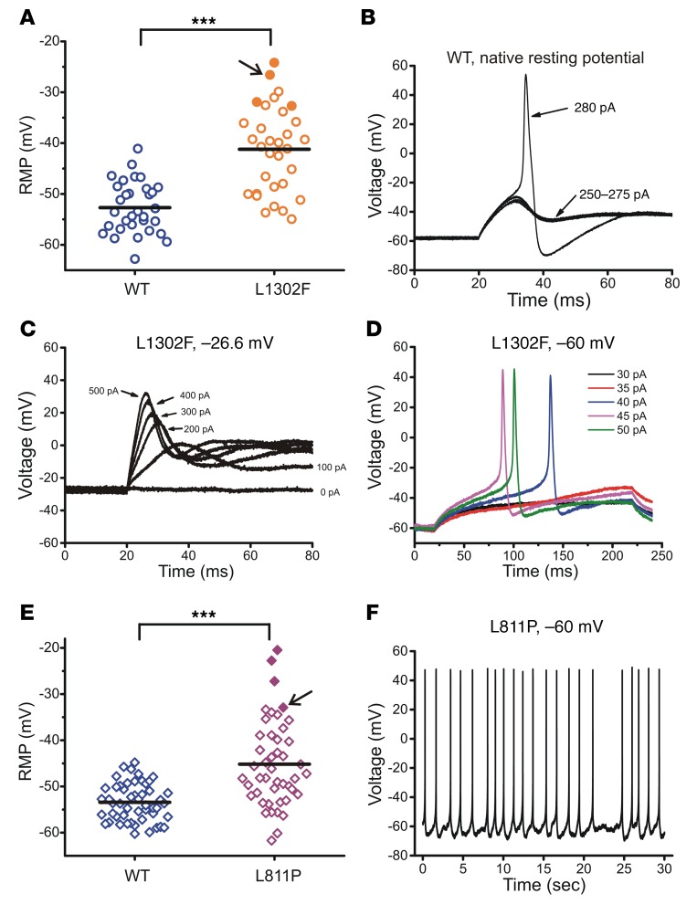
Figure 4. Effects of mutant channels on the RMP in small DRG neurons.
(A) Scatter-plot of the RMP recording from neurons expressing either WT or L1302F NaV1.9 channels. ***P < 0.001, by t test. Solid orange circles indicate cells that did not fire all-or-none action potentials in response to external stimuli at their native resting potentials (–24.2 mV, –26.6 mV, –31.9 mV, and –32.7 mV, respectively) but regained excitability when held at –60 mV. (B) Representative action potential in a small DRG neuron overexpressing WT channels evoked when the current injection reached 280 pA. The native resting potential for this neuron was –57.2 mV. (C) Small DRG neuron with a RMP of –26.6 mV (indicated by an arrow in panel A) did not fire action potentials in response to 200-ms current injections from 0 to 500 pA in 100-pA increments. (D) When held at –60 mV, the same neuron as that depicted in C produced subthreshold depolarizations in responses to 30-pA and 35-pA current injections and generated action potentials with a threshold current of 40 pA. (E) Scatter-plot of the RMP in neurons overexpressing either WT or L811P NaV1.9 channels. ***P < 0.001, by t test. Four cells indicated by solid purple diamonds did not fire action potentials in response to external stimuli at their native resting potentials (–20.5 mV, –22.7 mV, –27.2 mV, and –32.9 mV, respectively), but fired spontaneously when held at –60 mV (illustrated in panel F). The cell indicated by the arrow had a RMP of –32.9 mV. (F) Representative spontaneous firing from a holding potential of –60 mV recorded from the cell marked by an arrow in E.

