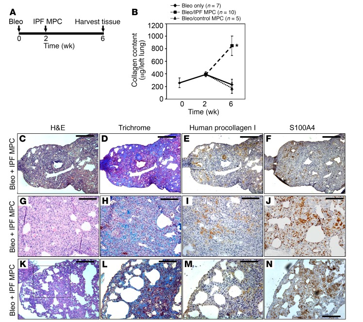
Figure 4. Human IPF MPCs convert the bleomycin model of lung fibrosis from a self-limited model to a model of persistent fibrosis.
(A) Schematic displaying timeline of mouse treatment protocol. (B) Three groups of immune-compromised mice were administered intratracheal bleomycin. In one group of mice (“bleo only”, n = 7), lungs were harvested at 2 and 6 weeks following bleomycin. In 2 groups of mice, 1 × 10⁶ IPF MPCs (n = 10 mice) or control MPCs (n = 5 mice) were injected via tail vein 2 weeks after bleomycin. Four weeks after administration of cells, lungs were harvested. Collagen content was quantified in left lungs by Sircol assay (data shown are aggregated from studies done using 2 independent IPF and control MPC lines). Data are expressed as mean ± SEM. *P < 0.01, by 2-tailed Student’s t test. (C–N) Serial 4-μm sections of right-lung tissue from mice receiving bleomycin followed by IPF MPCs. (C and D) Representative H&E and Trichrome stains assessing fibrosis and collagen distribution, respectively. Scale bar: 200 μm. (E and F) IHC using an antibody recognizing human procollagen to identify human cells and assess collagen synthesis (E) and an S100A4 antibody to assess the distribution of S100A4 and human procollagen–expressing cells (F). Boxed area in E showing procollagen+ human cells forming a fibrotic lesion obliterating an airspace. Scale bar: 200 μm. (G–J) Higher-power images demonstrating regions with new collagen deposition (G and H) heavily infiltrated with human cells expressing procollagen and cells expressing S100A4 (I and J). Scale bar: 100 μm. (K) Morphological analysis demonstrated the presence of peripheral fibrosis with cystic areas. Scale bar: 200 μm. (L–N) Trichrome stain (L) and IHC (M and N) of boxed region in K showing cystic change with heavy infiltration of procollagen+ human IPF MPCs codistributed with S100A4-expressing cells in fibrotic lesions. Scale bar: 100 μm.

