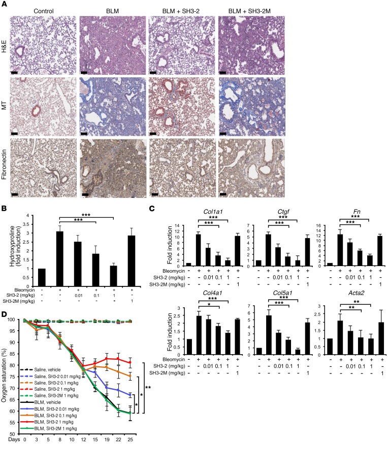
Figure 7. BLM-induced lung remodeling is attenuated by TAT–SH3-2.
(A) C57BL/6 mice were intratracheally treated with an equal volume of saline (control) or BLM (0.075 U). Beginning on day 14, mice were daily administered vehicle (METHOCEL/saline; control and BLM) or 1.0 mg/kg of the indicated TAT peptide (SH3-2 or SH3-2M) intraperitoneally. Lung tissue was harvested at day 28 and subjected to H&E staining for histology, Masson’s trichrome (MT) for collagen, or with anti-fibronectin and hematoxylin. Representative images from 5 animals are shown. Scale bars: 100 μm. (B) Mice were treated with saline or BLM as in A and beginning on day 14 administered daily vehicle (–) or the indicated concentration of TAT peptide. Animals were sacrificed on day 28 and hydroxyproline content determined as described in Methods. Data reflect mean ± SEM of n = 5. (C) Mice were treated as in B, and qPCR of the indicated genes assessed in lung tissue harvested on day 28. Data reflect mean ± SEM of n = 5. (D) Time-dependent fluctuation of oxygen saturation (SpO2) levels (determined on room air) in mice challenged with BLM (or saline) for 28 days and treated 1×/d with vehicle (METHOCEL/saline) or the indicated concentration of TAT–SH3-2 or TAT–SH3-2M beginning 14 days after initial BLM insult. Error bars reflect mean ± SEM from n = 5. *P < 0.05; **P < 0.005; ***P < 0.0005, 1-way ANOVA followed by Dunnett’s multiple comparisons test.

