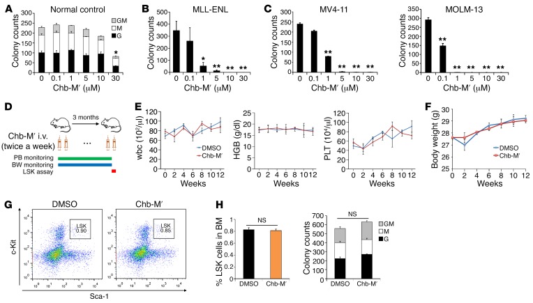
Figure 6. In vivo tolerability of Chb-M′.
(A) The number of granulocyte (G), macrophage (M), and G/M colonies were scored based on their morphology in a colony-forming cell assay. c-Kit+ bone marrow cells derived from wild-type C57BL/6 mice were cultured in the presence of indicated concentrations of Chb-M′ (n = 3). (B) The number of colonies was scored in a colony-forming cell assay with MLL-ENL–transduced mouse immortalized bone marrow cells in the presence of indicated concentrations of Chb-M′ (n = 3). (C) The number of colonies was scored in a colony-forming cell assay with MV4-11 or MOLM-13 cells in the presence of indicated concentrations of Chb-M′ (n = 3). (D) Schematic representation of treatment and monitoring schedule in wild-type C57BL/6 mice. PB, peripheral blood; BW, body weight. (E and F) Peripheral blood cell counts (E) and body weight (F) in mice receiving Chb-M′ or DMSO treatment for 3 months (n = 3). HGB, hemoglobin; PLT, platelets. (G and H) Frequency of LSK fraction was determined in bone marrow cells extracted from mice after a 3-month treatment with Chb-M′ or DMSO (n = 6). Representative FACS plots (G) and cumulative data (H) are shown. (I) The number of GM, G, and M colonies were scored based on their morphology in a colony-forming cell assay with c-Kit+ bone marrow cells extracted from mice after a 3-month treatment with Chb-M′ or DMSO (n = 6). Data are the mean ± SEM values. *P < 0.05, **P < 0.01, NS, not significant, by 2-tailed Student’s t test.

