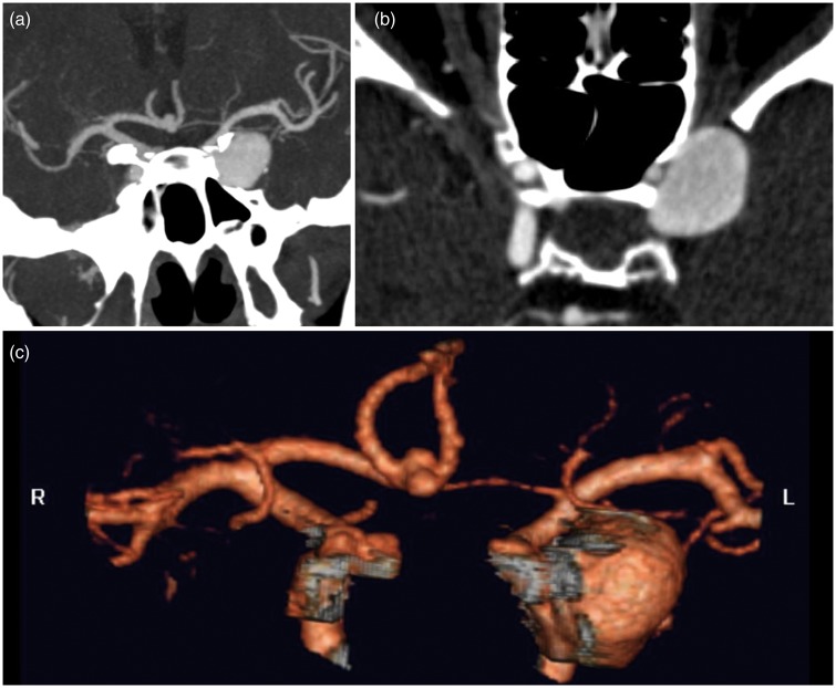
Figure 1.
(a) Computed tomography angiography shows multiple aneurysms located in the left internal carotid artery cavernous/ophthalmic segment (19 mm × 8 mm), right internal carotid artery cavernous segment (5 mm × 4 mm) and anterior communicating artery (4 mm × 2 mm). Notice the left internal carotid artery narrowing distally to the aneurysm on (b).

