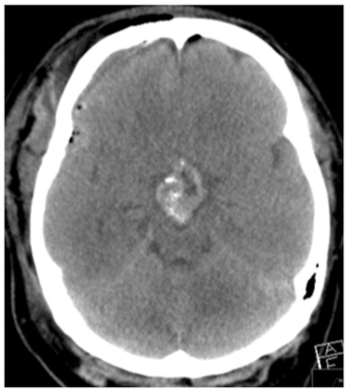
Figure 2.
The postoperative imaging showed a considerable decrease in the volume of tumour tissue. (a) T1-weighted sagittal and (b) T2-weighted coronal images, and no evidence of infarction. On the coronal T1-weighted sequence (c) haemorrhage was seen in the Sylvian fissure in the right and this was believed to be the cause of delayed spasm in our case.

