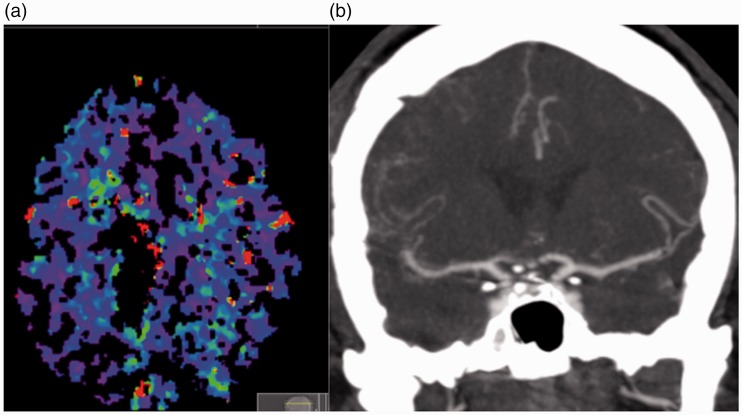
Figure 5.
A computed tomography (CT) perfusion scan (a) performed 24 hours after the stent-plasty procedure showed symmetrical perfusion (the image degradation was secondary to patient movement during the acquisition). A CT angiogram performed 7 days post-stent-plasty shows normal calibre to the M1 segment of the middle cerebral artery bilaterally.

