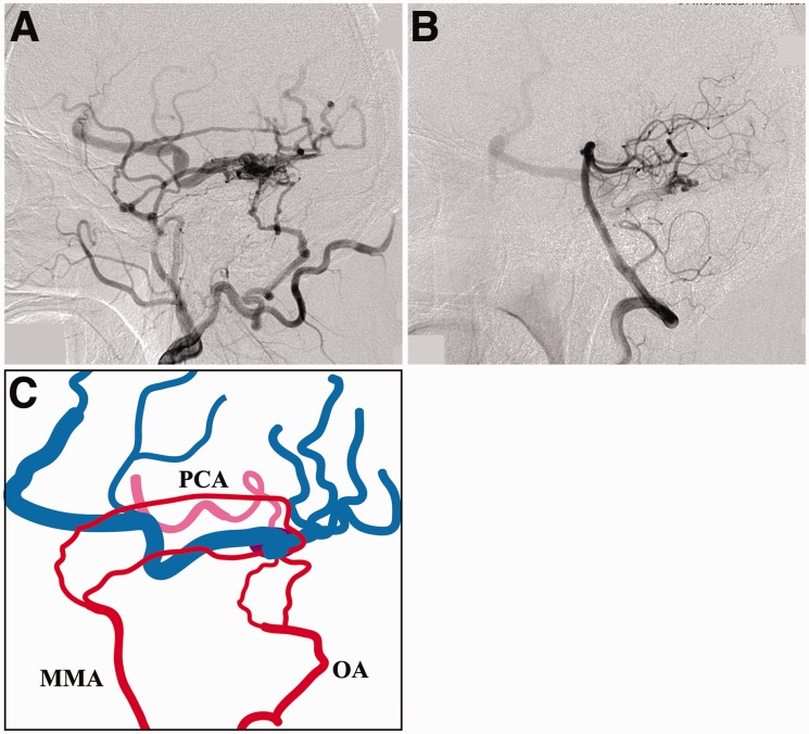
Figure 1.
a) The right external carotid artery angiogram demonstrating a tentorial dural arteriovenous fistula (DAVF). (b) The left vertebral artery angiogram. The DAVF is also fed by a pial branch originating from the right posterior cerebral artery (PCA). (c) Schematic drawing of the angioarchitecture showing the feeding arteries arising from the right external carotid artery (red) and from the PCA (pink), and the cortical venous drainage (blue). MMA: middle meningeal artery; OA: occipital artery; PCA: posterior cerebral artery.

