Figure 3.
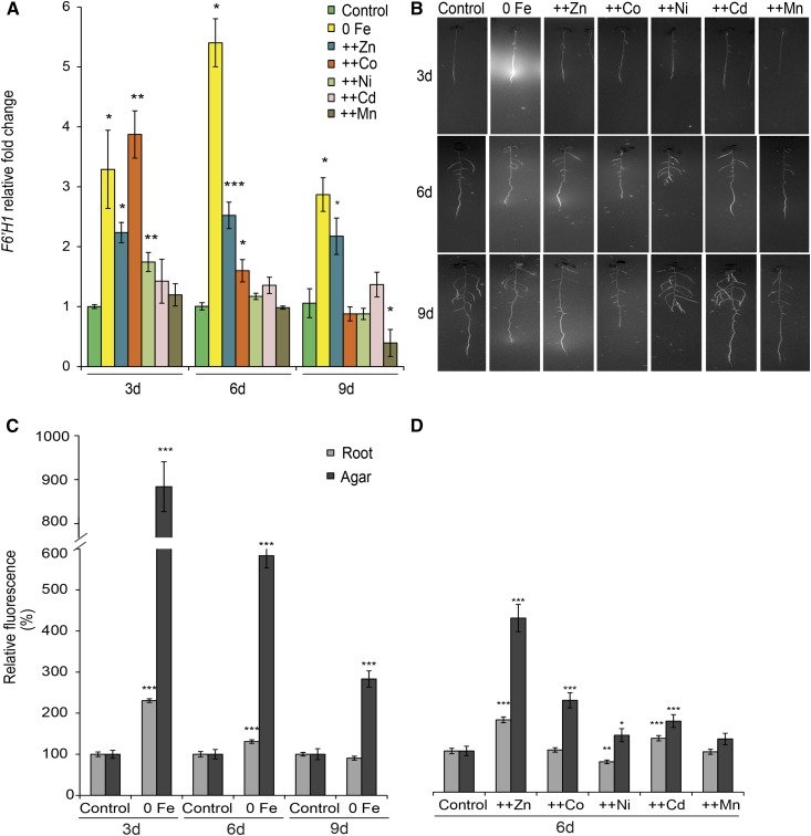
Effects of heavy metals on F6′H1 gene expression and the accumulation of fluorescent coumarins in roots and root exudates. A, Time course of F6′H1 transcript levels. B, UV fluorescence visualized at 365-nm wavelength. C and D, Relative fluorescence measured in roots and agar of plants grown on no supplementation of Fe + 50 µm ferrozine (0 Fe) for 3, 6, and 9 d (C) or excess heavy metal supplies for 6 d (D). Relative quantification was based on average pixel intensities of equally defined lengths of roots and agar portions around the roots. Seven-day-old seedlings grown on one-half-strength MS medium were transferred to one-half-strength MS medium with (control) or without Fe + 50 µm ferrozine (0 Fe) or with Fe and supplemented with 225 µm Zn (++Zn), 110 µm Co (++Co), 100 µm Ni (++Ni), 5 µm Cd (++Cd), or 1,500 µm Mn (++Mn). Bars represent means ± se; n = 25 to 30. Asterisks indicate statistically significant differences from controls at each time point according to Tukey’s test (*, P < 0.05; **, P < 0.01; and ***, P < 0.001).

