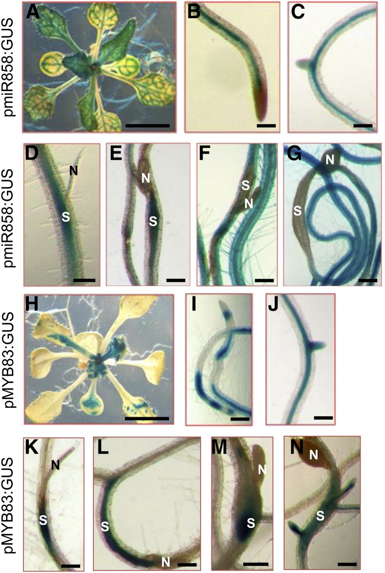
Figure 1.
Histochemical staining of GUS activity driven by miR858 and MYB83 promoters in transgenic Arabidopsis lines in response to H. schachtii infection. A to C, GUS activity of the pmiR858:GUS plants under noninfected conditions. Shown are GUS activity in the vascular tissues of leaves (A) and roots (B and C) of 2-week-old plants. D to G, GUS activity of the pmiR858:GUS plants in response to H. schachtii infection. Strong GUS activity was observed in the H. schachtii-induced syncytia at 3 (D) and 7 (E) dpi, whereas at 10 and 14 dpi, GUS activity was absent in the syncytia (F and G). H to J, GUS activity of the pMYB83:GUS plants under noninfected conditions. Shown are GUS activity in leaves (H) and vascular root tissues (I and J) of 2-week-old plants. K to N, GUS activity of the pMYB83:GUS plants in response to H. schachtii infection. Strong GUS activity was observed in the H. schachtii-induced syncytia at 3 (K), 7 (L), 10 (M), and 14 (N) dpi. N, Nematode; S, syncytium. Bars = 100 μm, except for A and H, which are 1 cm.

