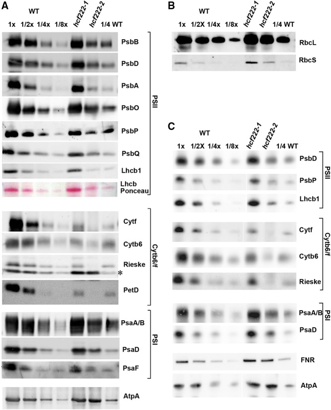
Figure 3.
Accumulation of chloroplast proteins in hcf222 mutants. A, Levels of chloroplast membrane proteins from seedlings cultivated for 20 d under moderate light conditions (40–60 μmol m−2 s−1). Whole-cell membranes from wild-type (WT) and mutant seedlings were separated by SDS-PAGE and transferred to nitrocellulose membranes. The dilution series contains 30 µg of protein (1x) or the indicated dilutions. Mutants contain 30 µg of membrane proteins. Antibodies used for detection are indicated on the right; the asterisk indicates an unspecific signal detected by the Rieske antibody. B, Levels of the large and small subunits of Rubisco (RbcL and RbcS) detected in whole-cell proteins of hcf222 mutants. C, Accumulation of thylakoid membrane proteins in hcf222 mutant seedlings grown under dim light conditions (5–10 μmol m−2 s−1). Plants were cultivated for 24 d. Whole-cell proteins were loaded on the gel. The dilution series in B and C contain 40 µg of protein (1x) or the indicated dilutions. Mutants contain 40 µg of whole-cell protein. Even if images were cut between lanes, all bands of a protein were detected simultaneously on one blot. Representative results of at least three biological replicates are depicted for each antibody.

