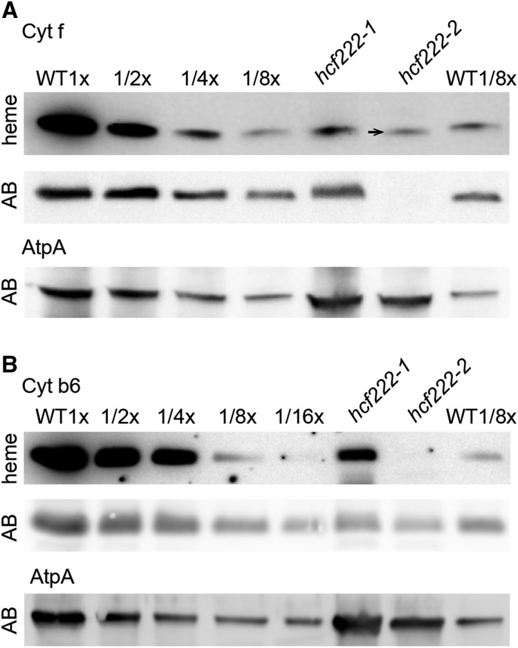
Figure 4.
Comparison of heme and protein levels in the wild type (WT) and hcf222 mutants. Covalently bound heme groups were detected by their peroxidase activity using luminol as a substrate. A, Detection of heme in cytochrome f. The arrow marks the slight shift of the heme signal in hcf222-2. Corresponding levels of the cytochrome f protein were detected with an antibody (panel AB). B, Heme detection in cytochrome b6 and the corresponding protein accumulation (gel AB). The AtpA subunit of the ATP synthase was used as a loading control. A total of 40 µg of protein (1x) or the indicated dilutions of the wild type was applied to the gel. The mutant lanes contained 40 µg of protein.

