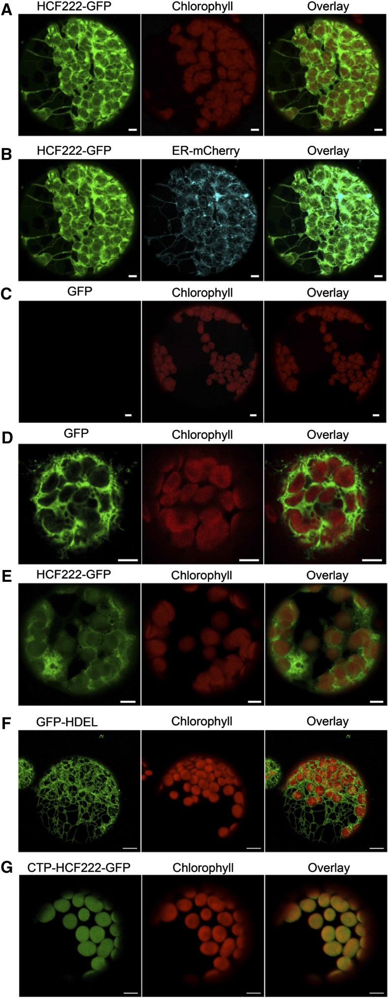
Figure 6.
Localization of HCF222 in the cell by confocal fluorescence microscopy. A, Analysis of N. benthamiana protoplasts transformed with HCF222-GFP. B, Visualization of the compartment marker ER-mCherry in relation to HCF222-GFP fluorescence (same protoplast as in A). C, Wild-type protoplast as a control, to verify that autofluorescence of chloroplasts did not influence GFP fluorescence (same microscope settings as in A and B). D, Control with cytosolic GFP. E, Visualization of HCF222-GFP fluorescence in Arabidopsis protoplasts that stably expressed the fusion protein in the mutant background of hcf222-2. F, Stably expressed compartment marker ER-GFP in the Arabidopsis line ER-gk (CS16251). G, Stable expression of CTP-HCF222-GFP in the mutant background of hcf222-2. Bars = 5 µm.

