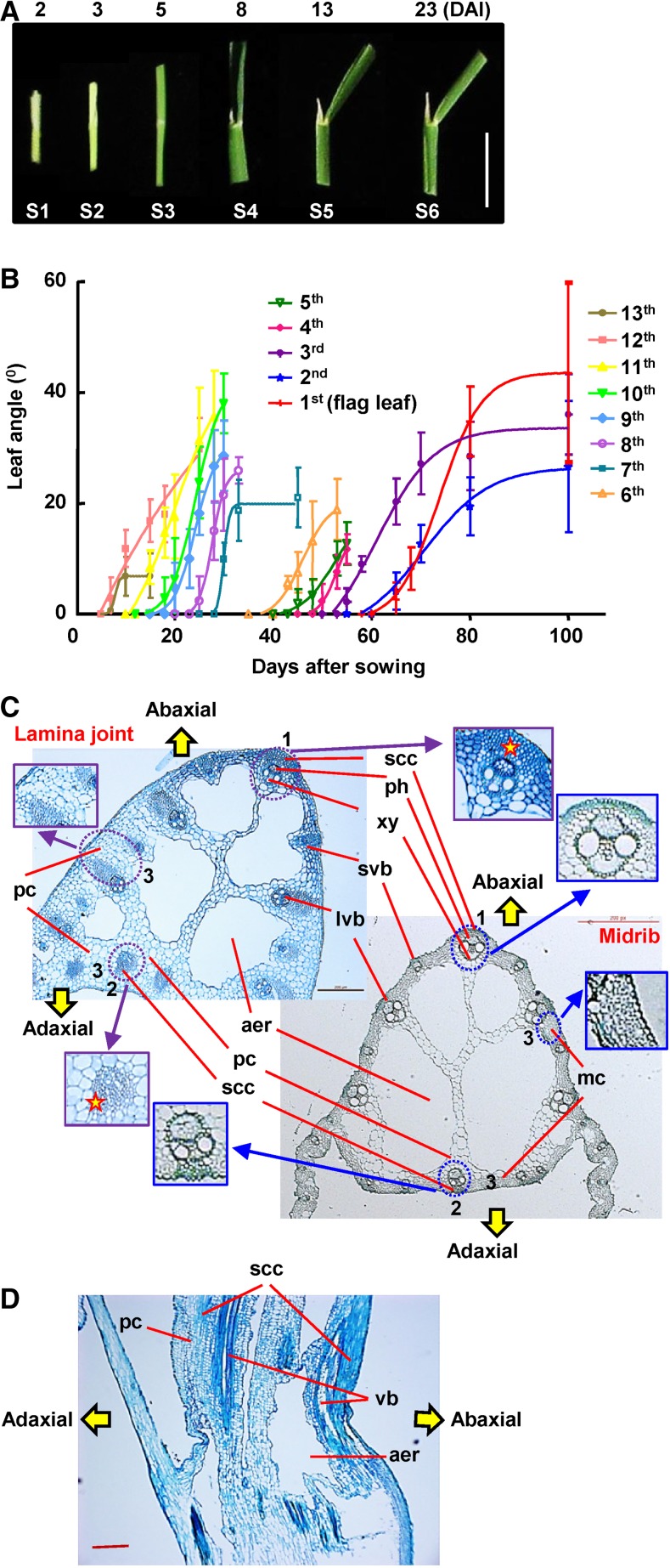
Figure 1.
Morphological and cytological observation of the rice lamina joint. A, Morphology of the lamina joint of rice flag leaf at 2, 3, 5, 8, 13, and 23 d (corresponding to stages 1–6) after lamina joint initiation (DAI). Representative images are shown. Bar = 1 cm. B, Statistics of lamina joint inclination of rice leaves. Angles (°) of each lamina joint at different days after sowing (DAS) were measured and statistically analyzed. Rice leaves from the bottom leaf (13th leaf, the first full leaf) to the flag leaf (first leaf) were analyzed. Experiments were biologically repeated three times, and data are shown as means ± sd (n > 10). C, Comparison of cytology between the lamina joint and midrib of mature leaf (the eighth leaf was used as representative) by cross-section analysis. Highlighted and magnified differences include layers and cell numbers of abaxial sclerenchyma cells (1) and adaxial sclerenchyma cells (2); the mesophyll cells in the midrib were replaced by multiple layers of large parenchyma cells in the lamina joint (3). aer, Aerenchyma; lvb, large vascular bundle; mc, mesophyll cell; pc, parenchyma cell; ph, phloem; scc, sclerenchyma cluster; svb, small vascular bundle; xy, xylem. Bar = 200 μm. D, Longitudinal section of the lamina joint of a mature flag leaf. vb, Vascular bundle. Bar = 200 μm.

