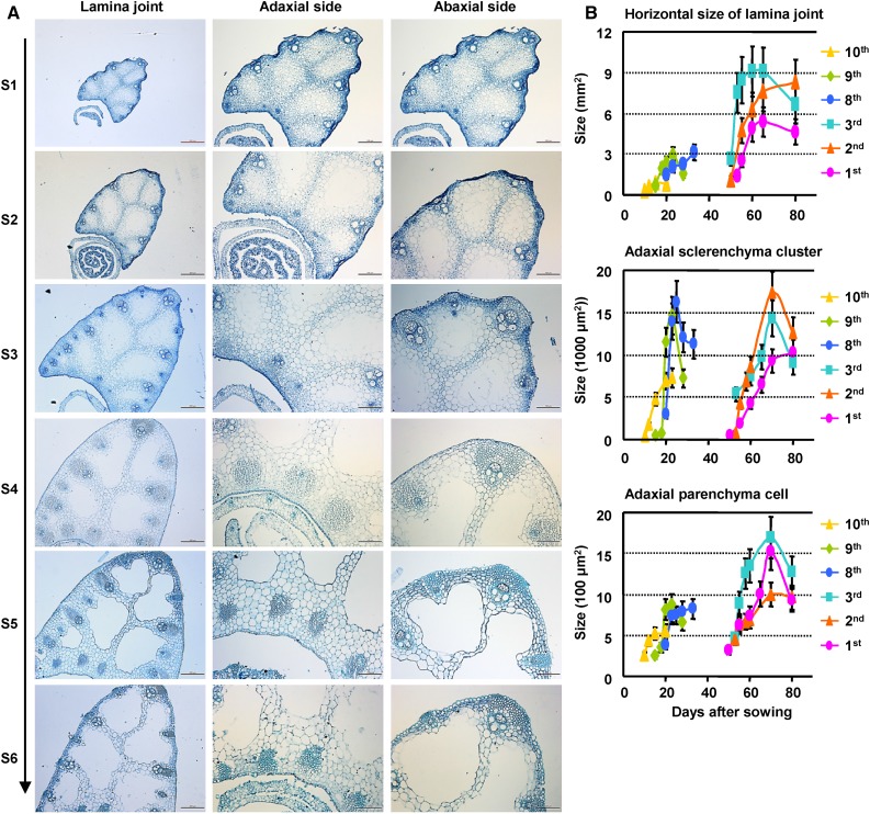
Figure 2.
Cytological dynamics of lamina joint development. A, Cytological dynamics of the lamina joint from initiation to maturation (left; bars = 200 μm), which can be divided into six stages (S1–S6). The lamina joint of the eighth leaf was observed by cross section, and the adaxial side (middle; bars = 100 μm) and the abaxial side (right; bars = 100 μm) were enlarged. B, Statistics of cytological changes of the lamina joint at different developmental stages. Lamina joints of first to third and eighth to 10th leaves were cross sectioned at different DAS, and sizes of horizontal area, adaxial sclerenchyma cluster, and adaxial parenchyma cells were measured and statistically analyzed. Data are shown as means ± sd (n = 3).

