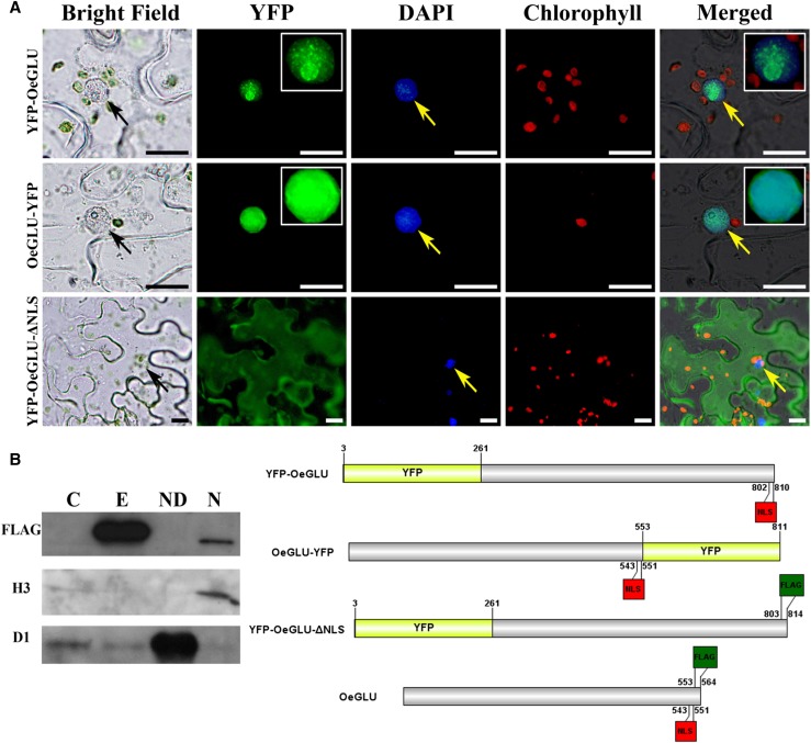
Figure 3.
OeGLU is localized in the nucleus. A, Epifluorescence microscopy of tobacco epidermal cells agroinfiltrated with YFP-fused constructs of OeGLU. OeGLU is localized in the nucleus as confirmed by both YFP-OeGLU and OeGLU-YFP constructs. Deletion of the predicted NLS (YFP-OeGLU-ΔNLS) resulted in a diffused pattern of fluorescence localized in the cytoplasm. Zoomed view (2×) of the nuclei of the YFP-OeGLU and OeGLU-YFP constructs under the YFP filter is depicted in insets. Nuclei were stained with DAPI. Scale bars, 10 μm. Arrows indicate nuclei. B, Western-blot analysis of crude protein extracts from tobacco plants agroinfiltrated with empty vector or with OeGLU. Nucleus-depleted and nuclear fractions of OeGLU. The protein samples were immunodetected either with anti-FLAG (sc-807), with antihistone H3 (nuclear marker), or with anti-D1 (chloroplastic marker) primary antibodies. Schematic representation of the constructs used. Numbers indicate the position of the amino acid residues. C, Empty vector; E, OeGLU; ND, nucleus-depleted; N, nuclear.

