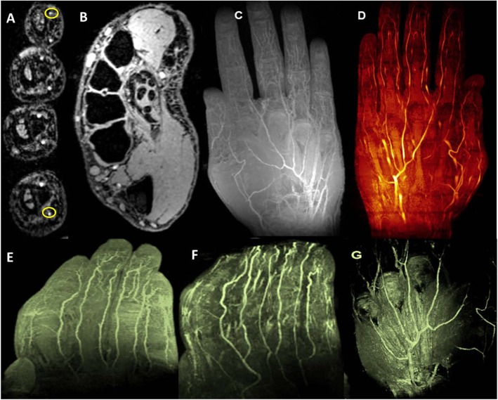
Fig 8.
7T TOF and T1 VIBE: A [TOF] demonstrates eight proper palmar digital arteries [two of them are yellow circled], digital tendons and synovial sheaths on axial view and B [T1 VIBE] shows proper palmer arteries and two proper palmer digital arteries in thumb, and cross-sectional transmetacarpal view highlighting intrinsic muscles, flexor digitorum superficialis and profundus tendons apparatus with synovial sheaths, ligamentous structures, and inter-metacarpal vasculature. C demonstrates multiple intensity projection [MIP] of hand vasculature and [D, E and F] represent non-contrast enhanced MR angiographic images of palmar and digital microvasculature in the hand. G demonstrates 3D view of volume rendering texture [VRT].

