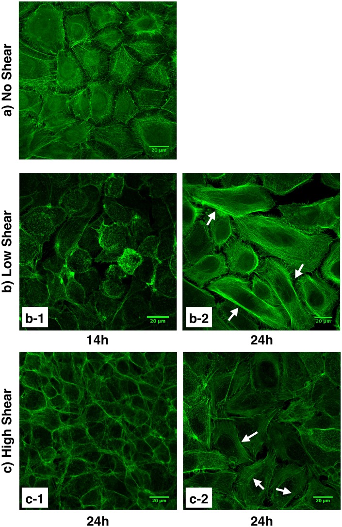
Fig 4. HCEC cytoskeleton organization.
Actin filaments were stained with fluorescently labeled phalloidin. a) Control cells (i.e., not exposed to shear stress). b) Cells exposed to low shear stress (4 dyn/cm2) for 14 hours (b-1) and 24 hours (b-2). Organization of the cytoskeleton with actin filament bundles are visible in cells exposed to low levels of shear stress for 24 hours. c) Cells exposed to high shear stress (8 dyn/cm2) for 24 hours with less visible (c-1) and more visible (c-2) filamentous actin cytoskeleton structure. White arrows indicate stretched actin filaments. Images captured using a Zeiss laser scanning confocal microscope.

