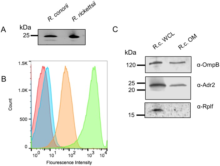
Fig 2. Outer-membrane expression of Adr2 in R. conorii.
(A) Western blot analysis using rabbit-anti-Adr2 antiserum confirms the expression in R. conorii Malish 7 and R. rickettsii Sheila Smith whole cell lysate (WCL). (B) Flow cytometry confirmed the expression of Adr2 at the surface of R. conorii. A shift in fluorescence is observed when fixed R. conorii was incubated with primary and secondary antibodies (orange) compared to samples prepared with only primary or secondary (red and blue, respectively). A sample is incubated with Anti-R. conorii serum (green) as a positive control of the flow cytometer. (C) Western blot analysis of R. conorii whole cell lysates (WCL) and outer-membrane(OM) preparations using antibodies against OmpB (mAb 6B6.6) or Adr2 demonstrates the presence of reactive species in both WCL and OM preparations. Anti-RplF (50s ribosomal protein L6) is used a control for cytoplasmic contents.

