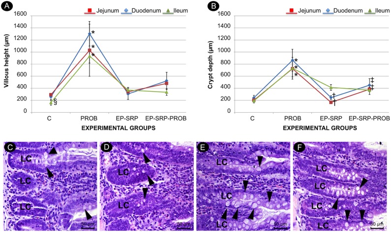
Fig 6. Histomorphometric analysis of small intestine.
Mean values and standard deviations of VH (A) and CD (B) in intestinal sections, with comparisons among groups. Photomicrographs of small intestine (duodenum sections): Group C (C); Group EP-SRP (D); Group PROB (E); Group EP-SRP-PROB (F). Abbreviations and symbols: LC = crypt of Lieberkühn; VH = villous height; CD = crypt depth; black arrowhead = calciform cells; * = Significant difference (p<0.05) when compared with Groups C, EP-SRP and EP-SRP-PROB; † = Significant difference (p<0.05) between Groups EP-SRP and EP-SRP-PROB; ‡ = Significant difference (p<0.05) between Groups C and EP-SRP-PROB; § = Significant difference (p<0.05) between Groups C and EP-SRP. Scale bars: C-F = 50 μm. (Hematoxylin and Eosin stain).

