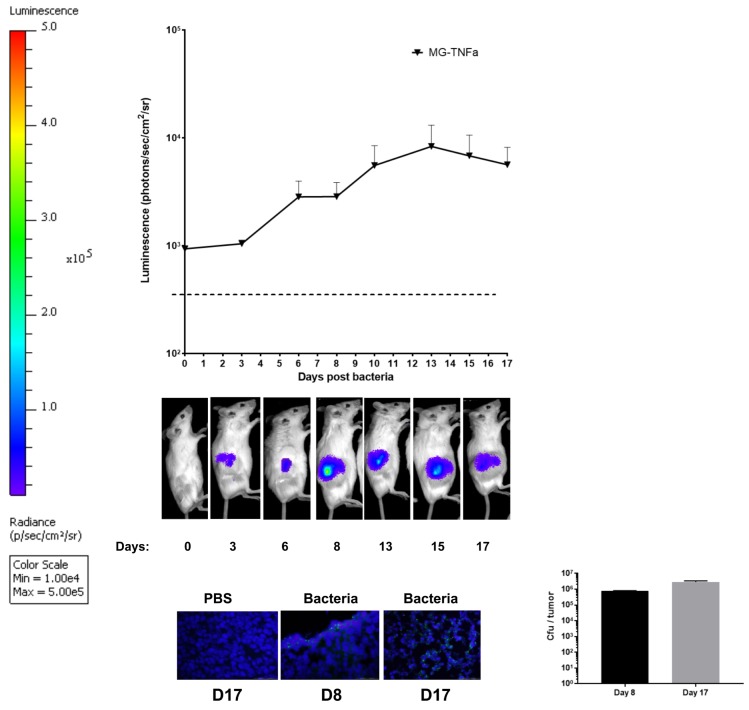
Fig 3. Intravenous administration of MG-Tnfα to tumour bearing mice.
Balb/C mice bearing s.c. CT26 flank tumours (n = 6) received 106 cfu of MG-TNFα i.v.. Growth of bacteria in tumours was analysed by (a) BLI and (b) immunofluorescence (IF), while (c) plating of tumour extracts on agar plates quantified viable bacteria. A representative image for each BLI group is shown. For IF, tissue sections from 2 individual mice per time point were analysed by fluorescence microscopy. (Original magnification, 400x), Scale bars, 50 μm.

