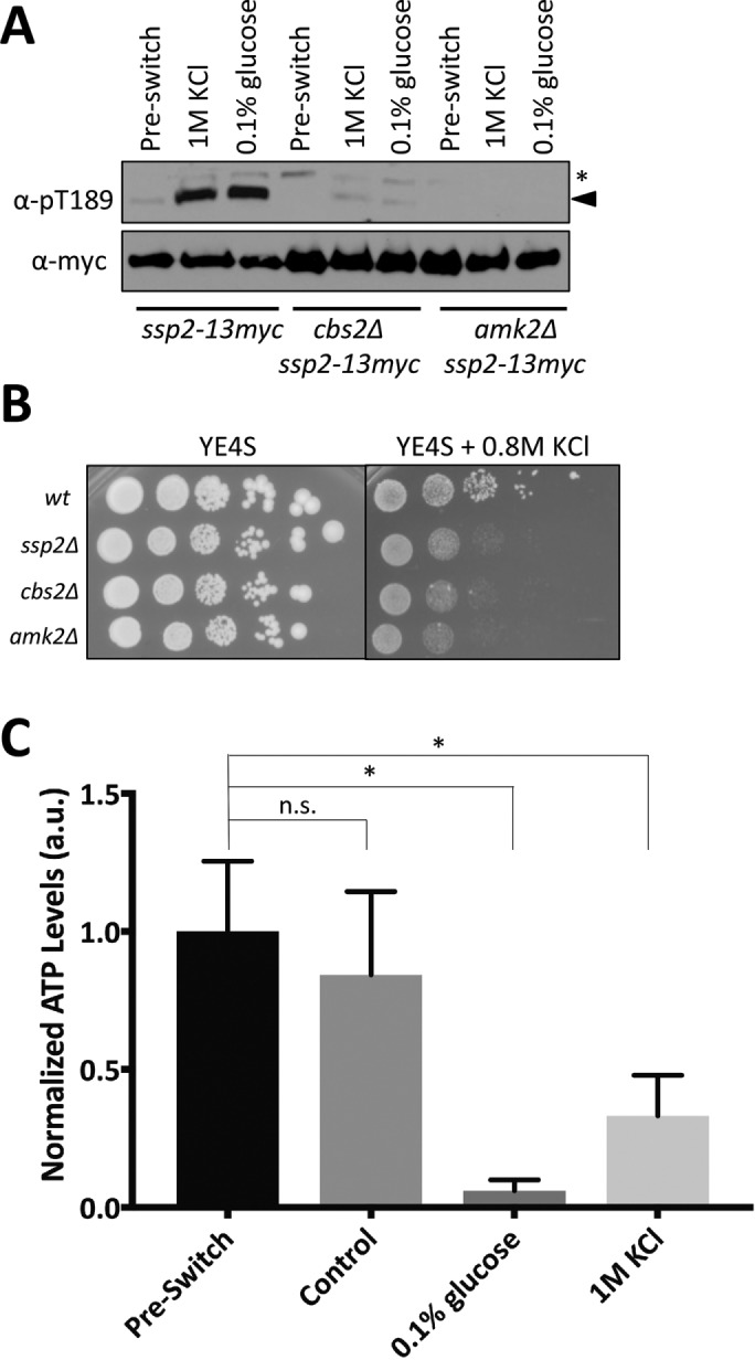
FIGURE 5:

AMPK senses depletion of cellular ATP levels during osmotic stress. (A) Western blot showing activation of Ssp2-pT189 in wild-type, cbs2∆, and amk2∆ cells in response to 1 M KCl. Preswitch (unstressed) and 0.1% glucose are used as a control for Ssp2 activation. Stress treatments were for 15 min. We used α-myc as a loading control for total Ssp2. For α-Ssp2-pT189, asterisks denote background bands, and arrowheads mark Ssp2-pT189 bands. (B) Tenfold serial dilutions of the indicated strains were spotted onto control (YE4S) plates or plates containing 0.8 M KCl. Cells were grown at 32°C. (C) Change in cellular ATP levels by 5-min treatment with the indicated stresses (preswitch and control were unstressed YE4S medium). ATP levels were measured and normalized as described in Materials and Methods. Mean ± SD. An unpaired t test was performed for statistical analyses, and p values were based on two-tailed distributions (*p < 0.05).
