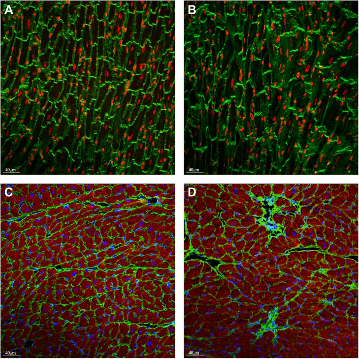
Fig 2. Gross cardiac morphology of hearts treated with doxorubicin.
Representative vinculin staining (green) in WT (PBS) mice (A) and WT doxorubicin (B). Nuclei (red) were visualized with draq5. Replacement fibrosis was detected in the doxorubicin treated hearts (D) but not in vehicle hearts (C), visualising with the anti-collagen VI antibody (green). Nuclei (blue) were visualised with DAPI, (E, F).

