Figure 2.
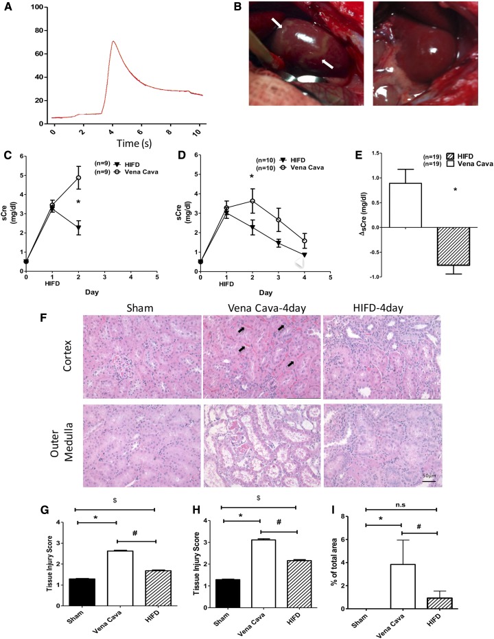
HIFD improves renal function and structure post-AKI. Rats were subjected to unilateral nephrectomy and unilateral I/R and allowed to recover for 24 hours before HIFD or vena cava saline infusion. (A) Representative tracing of renal venous pressure during HIFD, (B) (left panel) gross images of post-AKI kidney before, and (B) (right panel) 3 minutes after HIFD in the same kidney. Note elimination of striated zones of hypoperfusion post-HIFD (left panel, arrows). (C and D) Serum creatinine (sCre) versus time after HIFD or vena cava injection 24 hours post-I/R in rats recovering for 2 days (C) or 4 days (D) postsurgery. (E) Change in serum creatinine in the 24-hour period immediately after HIFD in all 2- and 4-day–treated rats (A–E); N for each group is shown at each time point; *P<0.05 in HIFD versus vena cava by unpaired t test. (F) Representative renal histology 4 days post-I/R. Cross sections of hematoxylin and eosin stained cortex and outer medulla from sham, I/R + vena cava injection, or I/R + HIFD in cortex are shown. Black arrows indicate congested RBCs in peritubular capillaries. (G and H) Renal injury scores on the basis of renal histology of kidney tissue ([G] cortex; [H] outer medulla) from (F). (I) Quantification of cortical vascular congestion expressed as percentage of total area from (F). (G–I) *,$P<0.05 for sham versus *I/R + vena cava or $I/R + HIFD; #P<0.05 for I/R + vena cava versus I/R + HIFD by paired t test. *P<0.05 in HIFD versus vena cava by unpaired t test.

