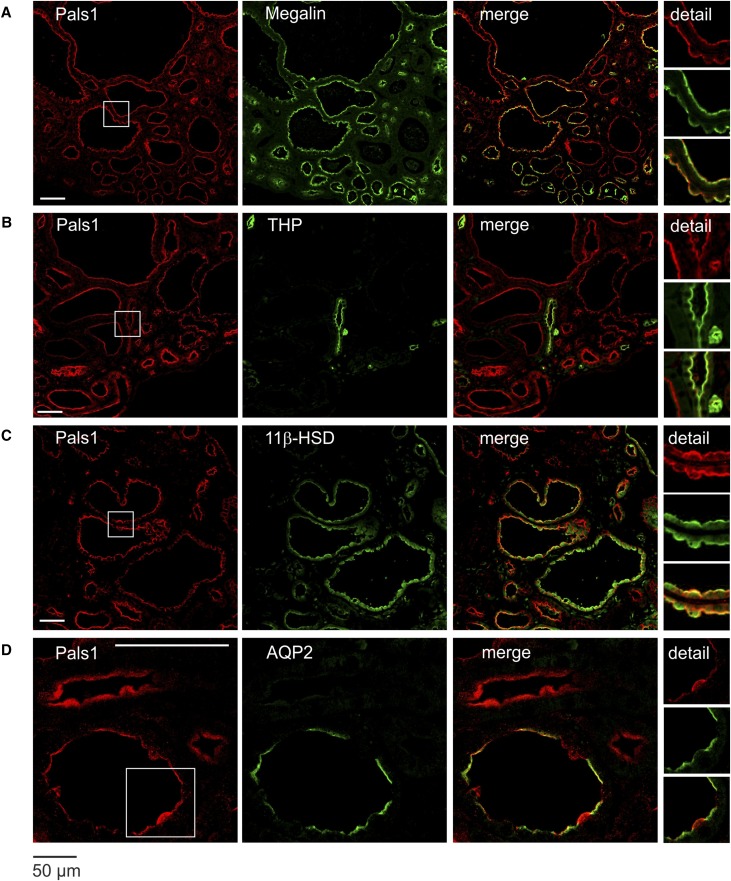
Figure 3.
Reduced Pals1 expression during nephrogenesis results in the formation of cysts. Immunofluorescence staining of Pals1 and costaining with diverse tubulus markers in Six2-Cre+ kidneys shows that remaining Pals1 is located to the apical membrane of all tubular cysts. Costaining of Pals1 with Megalin (proximal tubule marker, [A]), Tamm–Horsfall protein (thick ascending limb of the loop of Henle, [B]), 11β-HSD (distal tubules, [C]), and AQP2 (collecting duct, [D]). Scale bar, 50 µm.

