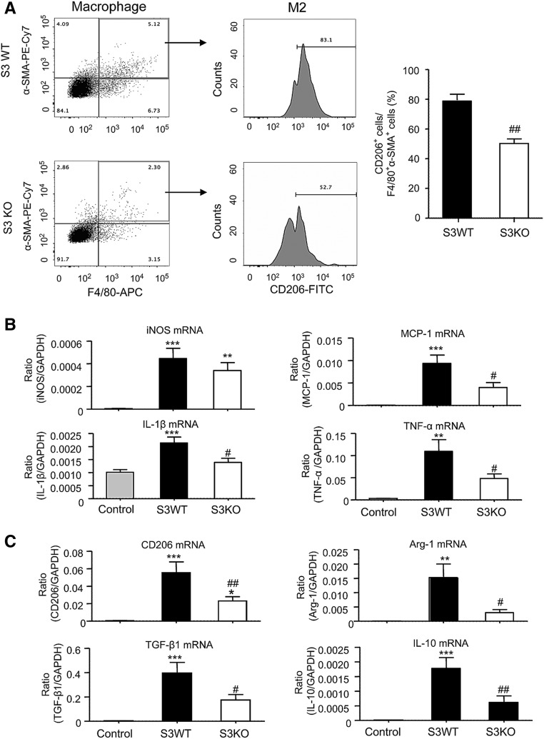
Figure 11.
Analysis of macrophage phenotype in grafted kidneys with chronic renal allograft rejection in Smad3 WT or KO recipient mice. Kidney allografts were transplanted into Smad3 WT or Smad3 KO recipient mice and examined 28 days later. An isograft group was used as a control. (A) Two-color flow cytometry analysis identifies F4/80+α-SMA+ MMT cells followed by analysis of CD206 expression by the gated MMT cells. Graph shows a significant reduction in the percentage of MMT cells expressing CD206 in renal allografts in Smad3 KO recipient mice. (B) Real-time PCR analysis of RNA extracted from whole renal allograft tissue for M1 macrophage markers (iNOS, MCP-1, IL-1β, and TNF-α). (C) Real-time PCR analysis for M2 macrophage markers (CD206, arginase-1, TGF-β1, and IL-10). Data are mean±SEM for groups of six mice. **P<0.01, ***P<0.001 versus isograft controls; #P<0.05, ##P<0.01 versus Smad3 WT mice.

