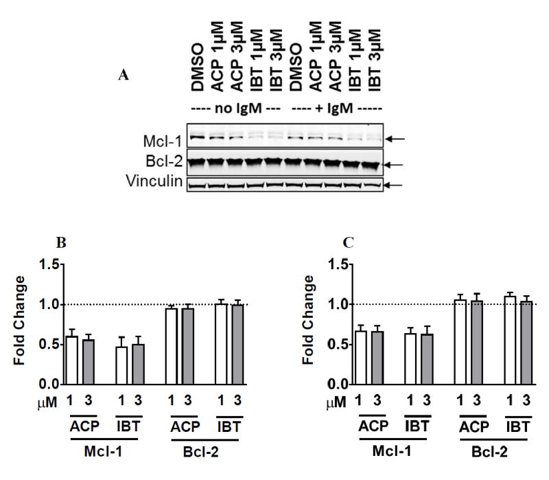
Figure 5. Comparison of ibrutinib and acalabrutinib-mediated changes in the levels of Bcl-2 and Mcl-1 antiapoptotic proteins.

A. CLL cells were incubated with DMSO only (controls) or with 1 μM or 3 μM ibrutinib or acalabrutinib for 48 hours without IgM stimulation (lanes 1-5) or with IgM stimulation (lanes 6-10). The protein lysates were immunoblotted for total Bcl-2 and Mcl-1 proteins. Vinculin was used as a loading control. B-C. Immunoblots were analyzed from five different patients, and changes in Mcl-1 and Bcl-2 in CLL cells treated with BTK inhibitors without (B) or with (C) IgM stimulation were quantitated. Values represent fold change relative to DMSO controls.
