Figure 2.

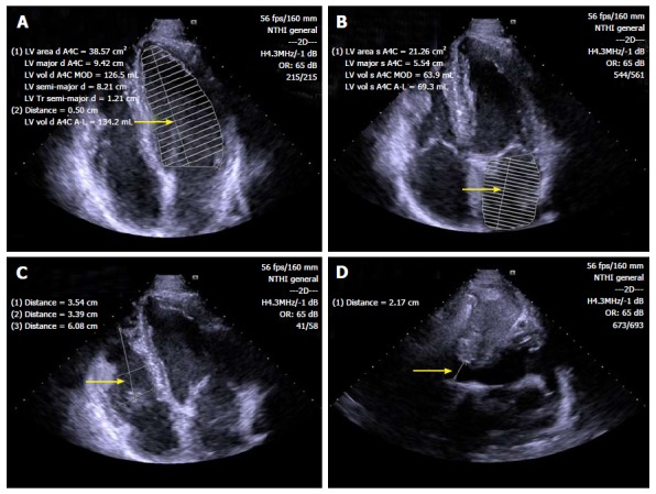
Standard B-mode echocardiography of endurance athlete showing enlargement of left ventricular (A), left atrial (B) and right ventricular (C) chambers, as well as inferior cavae vein dilatation (D) (arrows). LV: Left ventricular.

Standard B-mode echocardiography of endurance athlete showing enlargement of left ventricular (A), left atrial (B) and right ventricular (C) chambers, as well as inferior cavae vein dilatation (D) (arrows). LV: Left ventricular.