
Figure 1.
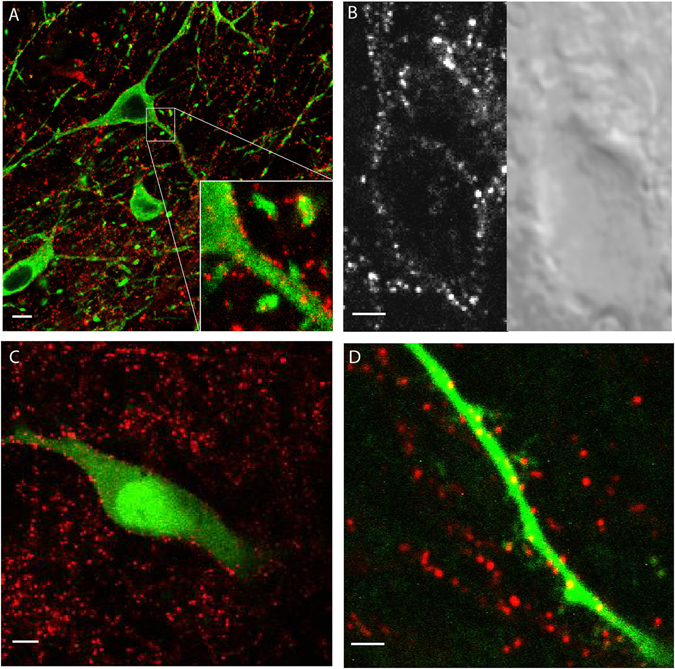
Visualizing the GFP-D2 receptor. (A) Confocal image of a slice from a GFP-D2 animal. Slice incubated in anti-GFP-AF594 (red) to amplify the signal on the D2 receptor prior to fixation. Tyrosine hydroxylase (TH) stained in green. (B) Simultaneously acquired 2-photon (left) and DIC (right) images of a live neuron from a GFP-D2 acute section. Slice was incubated in anti-GFP-AF594 prior to imaging. (C) 2-photon imaging of a live neuron filled with AF488. Slice was incubated in anti-GFP-AF594 prior to imaging. (D) 2-photon image zoomed in on a distal dendrite from a neuron filled with AF488. Receptors amplified with anti-GFP-AF594 (red) seen on dendritic shaft and spine-like structures. A-C scale bars 5 µm, D scale bar 1 µm.