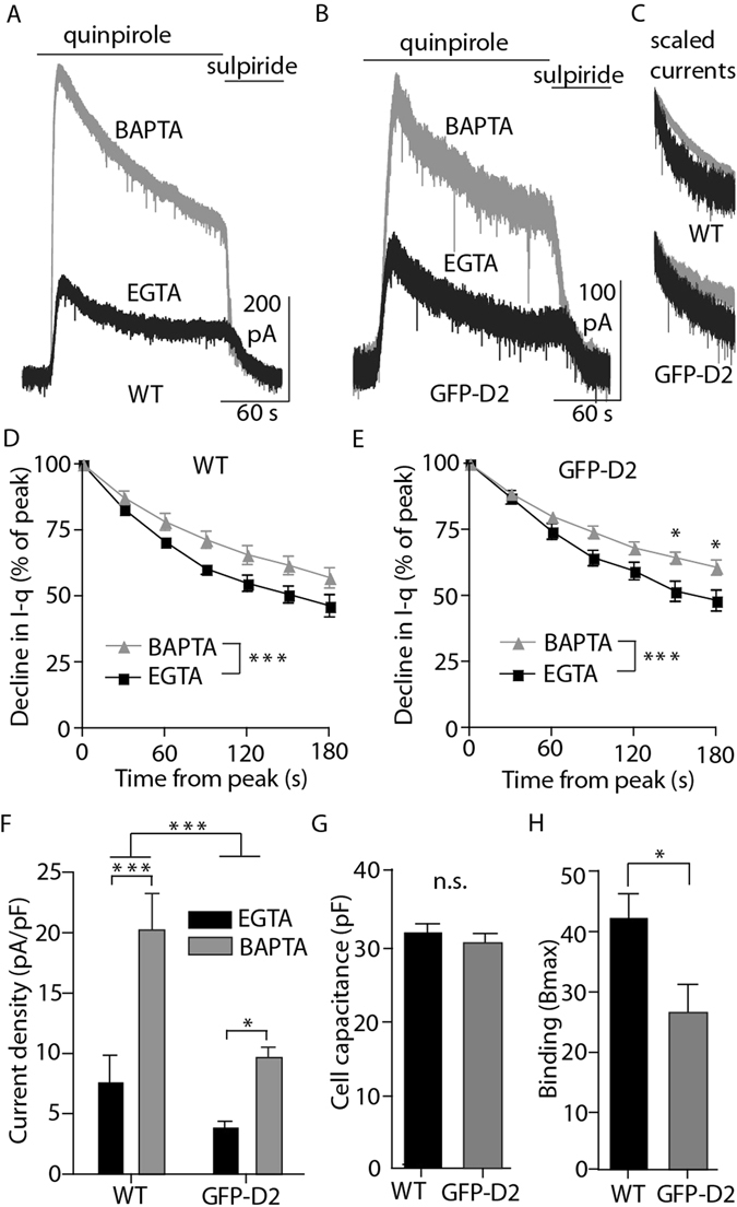
Figure 2.

Knocked-in D2 receptor functions in dopamine neurons, but is expressed at levels below the wild type. (A,B) Representative whole-cell voltage clamp recordings of dopamine neurons from wild type and GFP-D2 acute slices during the bath application of the D2 agonist quinpirole (10 µM) followed by the antagonist sulpiride (600 nM). (C) Example BAPTA and EGTA currents from wild type (top) and GFP-D2 (bottom) scaled and peak aligned. (D) In wild type, the quinpirole-induced current declines significantly more from peak value when EGTA internal is used compared to BAPTA (n = 5 and 7, two-way ANOVA followed by Bonferroni). (E) In the GFP-D2, the quinpirole-induced current also declines significantly more from peak value when EGTA internal is used compared to BAPTA (n = 7 and 10, two-way ANOVA followed by Bonferroni). (F) In both wild type and GFP-D2, the quinpirole-induced current density was larger using BAPTA internal compared with EGTA internal. As a group, wild type current density was larger than that for GFP-D2 (n = 5–10, two-way ANOVA followed by Bonferroni). (G) The average cell capacitance did not differ between the wild type and GFP-D2 groups (n = 21 and 29, unpaired t test). (H) The density of D2 receptors from midbrain samples as measured by the binding of [3H]YM-09151-2, was significantly greater in wild type compared to GFP-D2 (n = 9 each, unpaired t test). *p < 0.05, ***p < 0.001.
