
Figure 5.
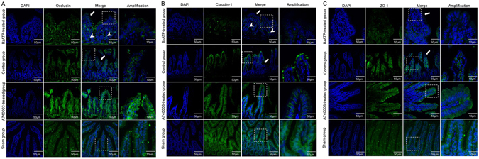
The effects of a P2X7R blockade on TJ distribution. Localization of occludin (A), claudin-1 (B), ZO-1 (C), and DAPI (DNA) within intestinal tissue sections assessed by immunofluorescence at 48 hours after CLP. TJ proteins (green) and DAPI stain (blue), merged TJ proteins and DAPI, as well as amplified merged TJ proteins and DAPI images are presented. Arrows indicate that biotin staining was ectopic to the lamina propria or deep within the epithelial surface. Arrowheads indicate the lack of focused staining on the surface of the villi.